Engineered Tissue Generates Its Own Capillaries
Researchers at Lawrence Livermore National Laboratory (LLNL) are investigating the ability to tissue-engineer compact 3D structures in order to accurately model the function of the central and peripheral nervous systems, the blood-brain barrier, the heart, and other human-body systems. These “in vitro Chip-based Human Investigational Platforms,” or iCHIPs, will provide a renewable source of human tissue for accurate and customizable in-vitro testing, replacing less-reliable test models such as animals. In the LLNL team’s most recent milestone, Monica Moya and Elizabeth Wheeler reveal a “bio-ink” that includes all of the necessary components to enable 3D-printed tissue to generate its own capillaries. They are currently answering questions regarding the project on Reddit.
Capillaries are permeable to certain molecules over chemical gradients. In the body, capillary beds cover the surfaces of individual organs and infiltrate cell matrices to perfuse nutrients from the bloodstream to the cells in the tissue. The body also has a natural hierarchical response to exercise and cell respiration, generating more capillaries where the demand for oxygen, calcium, and other nutrients is high. For this reason, vascularization, or the generation of blood vessels throughout the tissue, is critical for engineered tissue to be physiologically relevant and to stay alive for long periods of time.
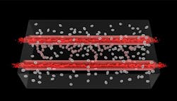
Using a 3D printer, the LLNL team introduces a streamlined, in-vitro, tissue-engineering process that layers a cellular substrate capable of supporting and growing a vascular system. The substrate includes cytoskeleton fibers, stem cells, and the necessary growth factors that enable differentiation from endothelial stem cells to adult vascular endothelial cells. This differentiation occurs later in the process.
The printer then builds large blood vessels that are incorporated into the substrate, at which point the team introduces blood flow through them. This acts as a stimulus, causing the growth factors in the tissue to activate sequences of the stem-cell DNA so that they differentiate into capillary cells. The tissue continues to build its own capillaries, bringing molecules from the blood supply to adjacent cells. The result is a 3D tissue with a vascular system.
“Bioprinting adds another dimension to tissue-on-a-chip platforms,” says Wheeler, principal investigator for iCHIP. “Having the ability to control the 3D structural environment, along with growing vessel networks to support the growing tissue, is one part of replicating the complexity of the human body.”
Now entering the third year of the project, Moya and Wheeler discuss methods to emulate the body's hierarchical ability to generate denser capillary beds in areas that need more oxygen and nutrients. So far, their strides in tissue self-vascularization should help improve the survival rate of cells in engineered tissue.
“Although printing implantable organs is not in the immediate future, our bio-printed tissue patches can be applied to toxicology studies, medical treatment testing and provide a test bed for fundamental science,” says Moya.
Watch the animation in the video below:
Moya and Wheeler are currently answering questions regarding the project on Reddit.
About the Author
Leah Scully
Associate Content Producer
Leah Scully is a graduate of The College of New Jersey. She has a BS degree in Biomedical Engineering with a mechanical specialization. Leah is responsible for Machine Design’s news items that cover industry trends, research, and applied science and engineering, along with product galleries. Visit her on Facebook, or view her profile on LinkedIn.
Voice Your Opinion!
To join the conversation, and become an exclusive member of Machine Design, create an account today!

Leaders relevant to this article: